|
 |
 |
| |
 |
|
| |
 |
 |
|
| |
|
|
| |
|
| |
|
| |
|
|
| |
|
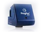
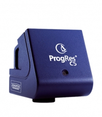
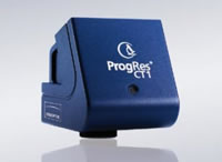
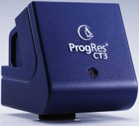
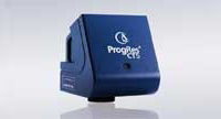
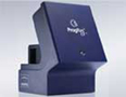
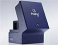
| ProgRes® CMOS Cameras CT1 / CT3 / CT5 |
 |
| Experience the high performance |
| |
 |
| Fast live image |
|
| Designed to provide highest versatility and cost-effective- ness, the cameras of the ProgRes® CMOS range allow for quick and precise setting of specimen and microscope, and hence provide comfortable operation. Fast live images meet the requirements of professionals, and the outstanding CMOS technology makes these ProgRes® cameras the first choice imaging solution for usage in education institutes and training labs |
| |
|
| High speed and high resolution |
|
The new generation of ProgRes® CMOS based USB 2.0 and FireWire cameras offers rapid image refresh rates of up to 90 fps@VGA. Resolutions from 1 up to 5 mega pixels allow for optimal performance in low as well as high magnifica- tions.
Due to the large pixel size of 5.2 μm2 the ProgRes® CT1 delivers high frame rates of 30 fps in full resolution and offers high sensitivity for best image quality.
|
Benefits • High frame rates • Good color reproduction • Free ProgRes® capture software for easy operation • Fit to any PC and microscope • Safe investment |
|
|
|
| ACCESSORIES |
|
|
|
|
| |
|
| |
|
| CF Series |
|
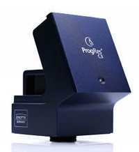
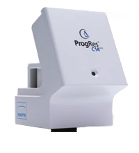
| ProgRes® CCD Research Cameras |
|
| Description |
|
Discover highest image quality
Highest image quality
The high sensitivity of the color and monochrome ProgRes® CCD Research cameras warrants brilliant images, especially when working with low-light specimens. Expeditious and smooth oper- ation is provided by sensitive CCD sensors with high frame rates and a broad dynamic range. Especially the cooled camera models are adapted to handle mainly low-noise long-time exposures.
ProgRes® CS / MS for highest sensitivity
Due to the very large pixel size of 8.3 μm these USB 2.0 cameras are especially suitable for most sensitive applications, where also a very fast live image is required. Even moving ob- jects can be recorded and fast processes can be followed up.
ProgRes® C14plus for true colors
For detailed image analysis and informative image docu- mentation, the Microscanning technology provided in the
scanning ProgRes® CCD Research cameras allows for capturing overview images and details in high-resolution of up to 12.5 mega pixels. The ProgRes® C14plus offers genuine color reproduction in proper detail with up to 12.5 mega pixels. Its patented Color-Co-Site-Sampling records the color information exactly in three color channels for an absolutely real color image.
Benefits ␣ High frame rates ␣ Perfect color reproduction ␣ Highest image resolution ␣ High sensitivity and low noise ␣ Fit to any PC and microscope ␣ Free ProgRes® capture software for easy operation ␣ Safe investment ␣ Excellent price-performance ratio |
|
|
|
| ACCESSORIES |
|
|
|
|
| |
|
| |
|
| SPEEDCORE SERIES |
|
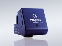
ProgRes® CCD SpeedXTcore
Cameras Reach your goal faster with SpeedXT core technology |
|
| Description |
|
Breakthrough in CCD speed
ProgRes® SpeedXT core 3 and SpeedXT core 5 are the first to feature Jenoptik’s innovative SpeedXT core technology providing very fast live speed rates of 17 fps / 13 fps in full resolution of 3 / 5 mega pixel. Due to the enhancement of the live image speed in combination with the high resolution the user is enabled to facilitate precise focusing and easy positioning of specimens without interlace effect in a more efficient way - a clear advantage in the analysis of moving objects and routine work in laboratories.
Exposure times up to 180 s ensure optimum captured im- ages, also under low-light conditions. The maximum possible color depth is 36 bit.
Superior color reproduction
An excellent color reproduction as well as ease of installation & operation are other distinguishing features of the cameras.
The software can be easily and quickly installed, enabling users to immediately capture brilliant images in excellent, acknowledged Jenoptik quality providing finest color gradings for sophisticated applications.
Benefits • SpeedXT core – outstanding CCD live image speed for
easy focussing • Excellent image quality and high resolution • Perfect color reproduction • ProgRes® Capture software for easy operation included • Easy and fast installation •
Excellent price-performance ratio
Reach your goal faster with SpeedXT core technology – faster installation, faster focussing, faster capture – in proven Jenoptik CCD quality! |
|
|
|
| ACCESSORIES |
|
|
|
|
| |
|
 |
|
| |
|
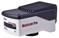
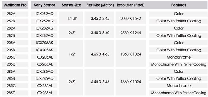
| MOTICAM PRO Series |
|
| New range of Motic CCD Scientific Cameras as a Complete or OEM package |
|
| Description |
|
| For many years, our unique "From Box to Pics" approach has given a wide range of markets unprecedented access to affordable Digital Microscopy. Our new Moticam Pro series take this approach to the CCD scientific level by offering as much as possible in a single box making this series our most powerful and flexible camera series yet. |
|
|
|
| |
|
|
|
 |
| |
| |
|
| |
|
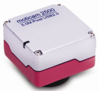
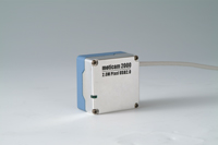
| MOTICAM 2500 |
|
Motic's newest Instant Digital Microscopy camera offers
an amazing 5 Megapixel live resolution. |
|
| Description |
|
The Moticam 2500 is a high-resolution live imaging microscopy camera.
On-chip software image enhancements include Noise-Reduction, Live Filtering and Settings Memory providing a constant environment for comparison work.
Motic Images Plus is an imaging suite included for both PC and Macintosh OSX platforms providing measurement, analysis and sharing tools for teachers, students and researchers alike.
With all necessary accessories included, the Moticam 2500 fits virtually any Microscope's eyepiece therefore allowing you to turn any microscope into a Digital Microscope. As an additional option, the Moticam package includes a B&S adapter to fit the camera directly into the microscope"s Eyetube therefore eliminating one lens assembly. |
|
|
|
| ACCESSORIES |
| |
|
|
|
| |
|
| |
|
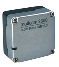
| MOTICAM 2300 |
|
| Motic's newest Instant Digital Microscopy camera offers a whopping 3 Megapixel live resolution. |
|
| Description |
|
The Moticam 2300 is a high-resolution live imaging microscopy camera.
On-chip software image enhancements include Noise-Reduction, Live Filtering and Settings Memory providing a constant environment for comparison work.
Motic Images Plus is an imaging suite included for both PC and Macintosh OSX platforms providing measurement, analysis and sharing tools for teachers, students and researchers alike.
With all necessary accessories included, the Moticam 2300 fits virtually any Microscope's eyepiece therefore allowing you to turn any microscope into a Digital Microscope. As an additional option, the Moticam 2000 package includes a B&S adapter to fit the camera directly into the microscope"s Eyetube therefore eliminating one lens assembly. |
|
|
|
| ACCESSORIES |
|
|
|
|
|
| |
|
| MOTICAM 2000 |
|
| Professionally convert any Microscope into a high-resolution Digital Microscope. |
|
| Description |
|
The Moticam 2000 is a high-resolution live imaging microscopy camera. With a live image of 1600x1280 pixels through a convenient USB2.0 connection, this metal housing camera provides a crisp and clear imaging stream for presentation, investigation and documentation.
On-chip software image enhancements include Noise-Reduction, Live Filtering and Settings Memory providing a constant environment for comparison work.
Motic Images Plus is an imaging suite included for both PC and Macintosh OSX platforms providing measurement, analysis and sharing tools for teachers, students and researchers alike.
With all necessary accessories included, the Moticam 2000 fits virtually any Microscope's eyepiece therefore allowing you to turn any microscope into a Digital Microscope. As an additional option, the Moticam 2000 package includes a B&S adapter to fit the camera directly into the microscope's Eyetube therefore eliminating one lens assembly. |
|
|
|
| ACCESSORIES |
| |
|
|
|
|
| |
|
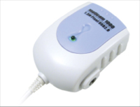
| MOTICAM 1000 |
|
| Affordable High-Resolution Live Imaging Camera |
|
| Description |
|
The Moticam 1000 is a competitively priced high-resolution live imaging microscopy camera. With a live image of 1280x1024 pixels, this image can be projected in any lecture or classroom environment without fear of pixelation. Enjoy crisp and clear live images.
Motic Images Plus is an imaging suite included for both PC and Macintosh OSX platforms providing measurement, analysis and sharing tools for teachers, students and researchers alike.
With all necessary accessories included, the Moticam 1000 fits virtually any Microscope's eyepiece therefore allowing you to turn any microscope into a Digital Microscope. |
|
|
|
| ACCESSORIES |
|
|
|
|
|
|
| |
|
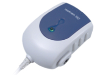
| MOTICAM 352 |
|
| An entry to professional and useful Digital Microscopy in an Educational or Hobby field. |
|
| Description |
|
| Use this camera with PC or Mac (OSX) systems and explore the Microscopic World with the included Motic Application Software Suite. Learn how to measure, edit and share captured video or still images or use the live video stream from the camera to teach an entire class |
|
|
|
| ACCESSORIES |
|
|
|
|
|
| |
|
| MOTICAM Image Plus |
|
| |
|
| Description |
|
| Motic Images Plus 2.0 is a Digital Microscopy Software Suite provided free of charge with most Motic Digital Microscopy items. Images Plus is available in Windows versions as well as OSX and contains powerful tools necessary in a wide range of applications from Educational to Professional Digital Microscopy. Users will find a familiar interface matched to either Windows or Macintosh OSX operating environments loaded with Measurment, Editing and Live Transmission features. This software package unlocks the potential of the microscope as a multi-media research, demonstration, hobby or analysis platform. With our own .sfc file format, users can easily create, send and edit multi-media images with voice narration and measurement tags. For those more forensic-minded, use the simple image comparison page to quickly compare images side by side. |
|
|
|
|
|
|
| |
|
| |
|
| MOTICAM Images Advanced 3.2 |
|
| Combined with Motic Images Assembly and Multi-Focus, this software Suite provides a professional digital microscopy analysis platform. |
|
| Description |
|
Motic Images Advanced has all of the features and tools of Motic Images Plus and much more. Use this software package to analyze fluorescence images and segment images by RGB or Gray Scale.
Motic Images Advanced is packaged with Motic's Multi-Focus and Assembly programs that will accurately allow you to assemble images in a vertical or horizontal platform taking into account any overlap or shifting of images. |
|
 |
PDF Brochure |
|
|
|
|
|
| |
|
| |
|
| MOTICAM Digilab II |
|
| DigiLab II is designed to work on exisiting Class C type local intranets. If your Lab, or Univeristy is already wired, then all you need is the software and you have DigiLab II installed. |
|
| Specifications |
|
The Teacher computer can:
- Have a live overview over up to 48 different live student microscopy images
- Have a live overview over up to 48 different live student screen images
- Take control of any student computer with mouse and keyboard control (this allows DigiLab II to be used as a cross-curriculum facilitator as other programs may also be controlled and manipulated through it)
- Launch and direct Digital Voice conversations
- Launch and direct Image and Text Chats
- Switch off, Reboot, Freeze, Logoff all of the student computers
- Present Live Measurements
- Broadcast Teacher's Microscope stream, Teacher's Desktop stream or any Student's Microscope image stream to the rest of the class
DigiLab II is also provided with Motic Images Advanced software for the teacher and Motic Images Plus 2.0 for the students
Please note that:
- Microsoft XP Pro (32-bit) is needed for this software
- Please check with your Motic supplier for additional guidlines, consultation and help for your individually tailored solution as some computer specifications are flexible and will reflect your choice in Digital Microscope or Moticam hardware.
|
|
|
|
|
|
|
| |
|
| |
|
| MOTIC Trace |
|
| Motic Trace is a software tool that helps turn any two microscopes into forensic comparison units. |
|
| Description |
|
Motic Trace is the software everyone has been waiting for. There is no need to buy expensive comparison microscopes. Simply connect two or more Moticams or Motic Digital Microscopes to your computer and get immediate live comparison imaging. Turn your existing microscopes into comparison scopes and break into forensic science teaching without breaking the bank.
Motic Trace will go further than existing Comparison Microscopes dare to go. Don't just compare images side by side. With this software you can overlay several live images, resize and independently rotate each of them. Match two fingerprints with ease from live images or recall from a saved set. Check handwriting authenticity in a matter of seconds or determine whether two pieces of fabric belong to the same set. Don't settle for expensive old technology, go Digital. |
| |
|
|
|
|
 |
|
| |
|
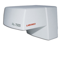
| iVU 7000 |
|
A perfect compliment to research
Ease of operation, super resolution and excellent color reproduction are the distinguishing characteristics of the iVu 7000 camera module. With 7.0 Megapixel resolution, the camera is the ideal tool for high-quality image documentation and elementary image analysis. |
|
| iVu 7000 Advantages: |
|
7.0 Megapixel image resolution with 20 frames per second (fps) at a high resolution, vivid color reproduction and high-fidelity in image contrast.
Working with still captured and live images is made easy with the use of ProgRes® CapturePro software. Elementary image analysis with fluorescene stains is also made easy with a host of software features.
|
|
|
|
| ACCESSORIES |
|
|
|
|
|
| |
|
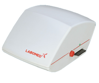
| iVU 5100 |
|
Digital, Video, and Secure Digital Capture...All In One
The super resolution iVu 5100 is a 5 Megapixel CMOS solution that boasts one of the fastest frame rates in its class, superb contrast, and unparalleled accuracy in color reproduction. This digital solution offers analog, digital, and an SD card interface in one highly affordable package. Total vertical integration of camera development at Labomed means better image quality, easy to navigate software, and better end-to-end software and driver support. |
|
| iVu 5100 Advantages: |
|
| 5 Megapixel resolution with 30 frames per second (fps), high-fidelity in image rendering and sharp image contrast. Users can stream live video, capture images and videos using PixelPro software, and capture images to an SD card. This system does it all!
PixelPro Image Capture Software makes still and video capturing simple. A modern and easy to navigate user interface allows users of all computer skills to quickly identify critical control functions, and to archive, measure and annotate images with complete ease. |
|
|
|
| ACCESSORIES |
|
|
|
|
|
| |
|
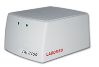
| iVU 3100 |
|
The iVu 3100 streamlines routine imaging and documentation tasks.
The iVu 3100 is a 3 Megapixel digital solution that boasts one of the fastest frame rates in its class, a high definition image, and unparalleled accuracy in color reproduction. Total vertical integration of camera development at Labomed means better image quality, easy to navigate software, and better end-to-end software and driver support. |
|
| iVu 3100 Advantages: |
|
3 Megapixel image resolution with 30 frames per second (fps), vivid color reproduction and high-fidelity in image contrast. Digital interfacing is accomplished through USB 2.0, making this system easy to integrate.
PixelPro Image Capture Software makes still and video capturing simple. A modern and easy to navigate user interface allows both teachers and students to quickly identify critical control functions, and to archive and annotate images with complete ease.
|
|
|
|
| ACCESSORIES |
|
|
|
|
| |
|
| |
|
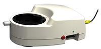
| iVU 55 |
|
A Versatile Imaging Solution for Medical Practices
The iVu S5 is a medical grade digital camera that can provide super resolution video and digital signals out, simultaneously. With an on-board SD Card slot, doctors need not be tethered to their computers to capture images for patient documentation. Consistent with Labomed's modular design philosophy, this camera will integrate seamlessly with your surgical microscope. |
|
| iVu S5 Advantages |
|
5 Megapixel resolution with 30 frames per second (fps), high-fidelity in image rendering and sharp image contrast. Digital, Analog, and SD Card capturing are a standard with the iVu S5. This system does it all!
PixelPro Image Capture Software makes still and video capturing simple. A modern and easy to navigate user interface allows users of all computer skills to quickly identify critical control functions, and to archive, measure and annotate images. |
|
|
|
| ACCESSORIES |
|
|
|
|
|
 |
|
| |
|
|
 |
|
| |
| Contact us for more details |
|
|
| |
|
|
|
| |
|
|
 |
|
|
|
 |
|